Gamma rays were discovered shortly after discovery of X-rays. In 1896, French scientist Henri Becquerel discovered that uranium minerals could expose a photographic plate through another material. Periodic Table
Discovery of Gamma Rays
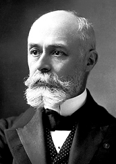
Antoine Henri Becquerel
Gamma rays were discovered shortly after discovery of X-rays. In 1896, French scientist Henri Becquerel discovered that uranium minerals could expose a photographic plate through another material. Becquerel presumed that uranium emitted some invisible light similar to X-rays, which were recently discovered by W.C.Roentgen. He called it “metallic phosphorescence”. In fact, Henri Becquerel had found gamma radiation being emitted by radioisotope 226Ra (radium), which is part of the Uranium series of uranium decay chain.Gamma rays were first thought to be particles with mass, for example extremely energetic beta particles. This opinion failed, because this radiation cannot be deflected by a magnetic field, what indicated they had no charge. In 1914, gamma rays were observed to be reflected from crystal surfaces, proving they must be electromagnetic radiation, but with higher energy (higher frequency and shorter wavelengths).
See also:
Description of Gamma Rays
See also:
Gamma Ray
See also:
Characteristics of Gamma Rays
We hope, this article, Discovery of Gamma Rays / Radiation, helps you. If so, give us a like in the sidebar. Main purpose of this website is to help the public to learn some interesting and important information about radiation and dosimeters.
Gamma rays, also known as gamma radiation, refers to electromagnetic radiation (no rest mass, no charge) of a very high energies. Definition of Gamma rays. Periodic Table
Gamma rays, also known as gamma radiation, refers to electromagnetic radiation (no rest mass, no charge) of a very high energies. Gamma rays are high-energy photons with very short wavelengths and thus very high frequency. Since the gamma rays are in substance only a very high-energy photons, they are very penetrating matter and are thus biologically hazardous. Gamma rays can travel thousands of feet in air and can easily pass through the human body.Gamma rays are emitted by unstable nuclei in their transition from a high energy state to a lower state known as gamma decay. In most practical laboratory sources, the excited nuclear states are created in the decay of a parent radionuclide, therefore a gamma decay typically accompanies other forms of decay, such as alpha or beta decay.Radiation and also gamma rays are all around us. In, around, and above the world we live in. It is a part of our natural world that has been here since the birth of our planet. Natural sources of gamma rays on Earth are inter alia gamma rays from naturally occurring radionuclides, particularly potassium-40. Potasium-40 is a radioactive isotope of potassium which has a very long half-life of 1.251×10⁹ years (comparable to the age of Earth). This isotope can be found in soil, water also in meat and bananas. This is not the only example of natural source of gamma rays.
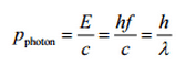
Photon
A photon, the quantum of electromagnetic radiation, is an elementary particle, which is the force carrier of the electromagnetic force. The modern photon concept was developed (1905) by Albert Einstein to explain of the photoelectric effect, in which he proposed the existence of discrete energy packets during the transmission of light.Before Albert Einstein, notably the German physicist Max Planck had prepared the way for the concept by explaining that objects that emit and absorb light do so only in amounts of energy that are quantized, that means every change of energy can occur only by certain particular discrete amounts and the object cannot change energy in any arbitrary way. The concept of modern photon came into general use after the physicist Arthur H. Compton demonstrated (1923) the corpuscular nature of X-rays. This was the validation that Einstein’s hypothesis that light itself is quantized.The term photon comes from Greek phōtos, “light” and a photon is usually denoted by the symbol γ (gamma). The photons are also symbolized by hν (in chemistry and optical engineering), where h is Planck’s constant and the Greek letter ν (nu) is the photon’s frequency. The radiation frequency is key parameter of all photons, because it determines the energy of a photon. Photons are categorized according to the energies from low-energy radio waves and infrared radiation, through visible light, to high-energy X-rays and gamma rays.Photons are gauge bosons for electromagnetism, having no electric charge or rest mass and one unit of spin. Common to all photons is the speed of light, the universal constant of physics. In empty space, the photon moves at c (the speed of light – 299 792 458 metres per second).
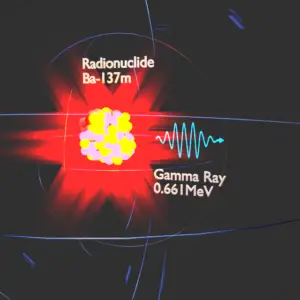
[/su_accordion]Barium-137m is a product of a common fission product – Caesium – 137. The main gamma ray of Barium-137m is 661keV photon.
See above:See also:
Gamma Ray
See also:
Discovery of Gamma Rays
We hope, this article, Description of Gamma Ray, helps you. If so, give us a like in the sidebar. Main purpose of this website is to help the public to learn some interesting and important information about radiation and dosimeters.
Gamma rays are electromagnetic radiation. Key features of gamma rays are summarized in following few points. Characteristics of Gamma Rays. Periodic Table
Characteristics of Gamma Rays / Radiation
Key features of gamma rays are summarized in following few points:
Gamma rays are high-energy photons (about 10 000 times as much energy as the visible photons),
The same photons as the photons forming the visible range of the electromagnetic spectrum – light.
Photons (gamma rays and X-rays) can ionize atoms directly (despite they are electrically neutral) through the Photoelectric effect and the Compton effect, but secondary (indirect) ionization is much more significant.
Gamma rays ionize matter primarily via indirect ionization.
Although a large number of possible interactions are known, there are three key interaction mechanisms with matter.
Gamma rays travel at the speed of light and they can travel thousands of meters in air before spending their energy.
Since the gamma radiation is very penetrating matter, it must be shielded by very dense materials, such as lead or uranium.
The distinction between X-rays and gamma rays is not so simple and has changed in recent decades. According to the currently valid definition, X-rays are emitted by electrons outside the nucleus, while gamma rays are emitted by the nucleus.
We hope, this article, Characteristics of Gamma Rays / Radiation, helps you. If so, give us a like in the sidebar. Main purpose of this website is to help the public to learn some interesting and important information about radiation and dosimeters.
Although a large number of possible interactions of gamma radiation with matter are known, there are three key interaction mechanisms with matter. Periodic Table
Description of Gamma Radiation
Gamma rays, also known as gamma radiation, refers to electromagnetic radiation (no rest mass, no charge) of a very high energies. Gamma rays are high-energy photons with very short wavelengths and thus very high frequency. Since the gamma rays are in substance only a very high-energy photons, they are very penetrating matter and are thus biologically hazardous. Gamma rays can travel thousands of feet in air and can easily pass through the human body.Gamma rays are emitted by unstable nuclei in their transition from a high energy state to a lower state known as gamma decay. In most practical laboratory sources, the excited nuclear states are created in the decay of a parent radionuclide, therefore a gamma decay typically accompanies other forms of decay, such as alpha or beta decay.Radiation and also gamma rays are all around us. In, around, and above the world we live in. It is a part of our natural world that has been here since the birth of our planet. Natural sources of gamma rays on Earth are inter alia gamma rays from naturally occurring radionuclides, particularly potassium-40. Potassium-40 is a radioactive isotope of potassium which has a very long half-life of 1.251×10⁹ years (comparable to the age of Earth). This isotope can be found in soil, water also in meat and bananas. This is not the only example of natural source of gamma rays.
Barium-137m is a product of a common fission product – Caesium – 137. The main gamma ray of Barium-137m is 661keV photon.
Characteristics of Gamma Rays / Radiation
Key features of gamma rays are summarized in following few points:
Gamma rays are high-energy photons (about 10 000 times as much energy as the visible photons), the same photons as the photons forming the visible range of the electromagnetic spectrum – light.
Photons (gamma rays and X-rays) can ionize atoms directly (despite they are electrically neutral) through the Photoelectric effect and the Compton effect, but secondary (indirect) ionization is much more significant.
Gamma rays ionize matter primarily via indirect ionization.
Although a large number of possible interactions are known, there are three key interaction mechanisms with matter.
Gamma rays travel at the speed of light and they can travel thousands of meters in air before spending their energy.
Since the gamma radiation is very penetrating matter, it must be shielded by very dense materials, such as lead or uranium.
The distinction between X-rays and gamma rays is not so simple and has changed in recent decades. According to the currently valid definition, X-rays are emitted by electrons outside the nucleus, while gamma rays are emitted by the nucleus.
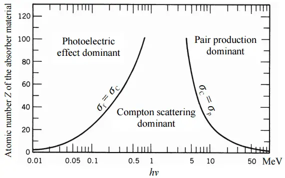
Image: The relative importance of various processes of gamma radiation interactions with matter.
The relative importance of various processes of gamma radiation interaction with matter.
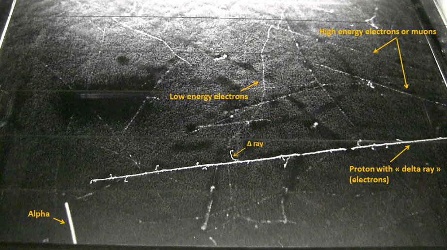
Comparison of particles in a cloud chamber. Source: wikipedia.orgTotal photon cross sections. Source: Wikimedia Commons
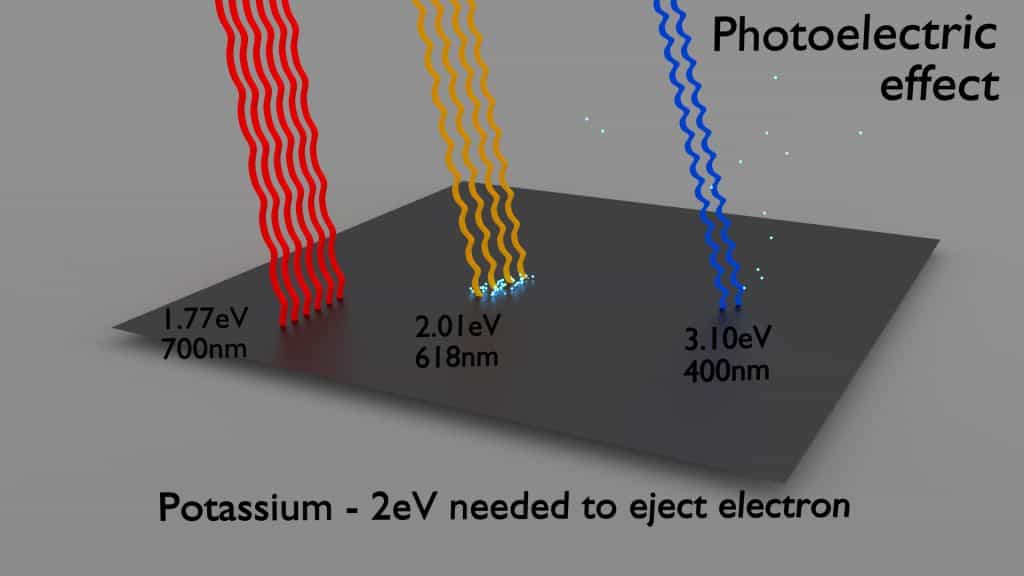
Photoelectric Effect
The photoelectric effect dominates at low-energies of gamma rays.
The photoelectric effect leads to the emission of photoelectrons from matter when light (photons) shines upon them.
The maximum energy an electron can receive in any one interaction is hν.
Electrons are only emitted by the photoelectric effect if photon reaches or exceeds a threshold energy.
A free electron (e.g. from atomic cloud) cannot absorb entire energy of the incident photon. This is a result of the need to conserve both momentum and energy.
The cross-section for the emission of n=1 (K-shell) photoelectrons is higher than that of n=2 (L-shell) photoelectrons. This is a result of the need to conserve momentum and energy.
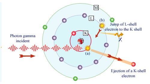
In the photoelectric effect, a photon undergoes an interaction with an electron which is bound in an atom. In this interaction the incident photon completely disappears and an energetic photoelectron is ejected by the atom from one of its bound shells. The kinetic energy of the ejected photoelectron (Ee) is equal to the incident photon energy (hν) minus the binding energy of the photoelectron in its original shell (Eb).
Ee=hν-Eb
Therefore photoelectrons are only emitted by the photoelectric effect if photon reaches or exceeds a threshold energy – the binding energy of the electron – the work function of the material. For gamma rays with energies of more than hundreds keV, the photoelectron carries off the majority of the incident photon energy – hν.
Following a photoelectric interaction, an ionized absorber atom is created with a vacancy in one of its bound shells. This vacancy is will be quickly filled by an electron from a shell with a lower binding energy (other shells) or through capture of a free electron from the material. The rearrangement of electrons from other shells creates another vacancy, which, in turn, is filled by an electron from an even lower binding energy shell. Therefore a cascade of more characteristic X-rays can be also generated. The probability of characteristic x-ray emission decreases as the atomic number of the absorber decreases. Sometimes , the emission of an Auger electron occurs.
Photoelectric effect with photons from visible spectrum on potassium plate – threshold energy – 2eVGamma absorption by an atom. Source: laradioactivite.com/
Cross-Sections of Photoelectric Effect
At small values of gamma ray energy the photoelectric effect dominates. The mechanism is also enhaced for materials of high atomic number Z. It is not simple to derive analytic expression for the probability of photoelectric absorption of gamma ray per atom over all ranges of gamma ray energies. The probability of photoelectric absorption per unit mass is approximately proportional to:
τ(photoelectric) = constant x ZN/E3.5
where Z is the atomic number, the exponent n varies between 4 and 5. E is the energy of the incident photon. The proportionality to higher powers of the atomic number Z is the main reason for using of high Z materials, such as lead or depleted uranium in gamma ray shields.
Although the probability of the photoelectric absorption of gamma photon decreases, in general, with increasing photon energy, there are sharp discontinuities in the cross-section curve. These are called “absoption edges” and they correspond to the binding energies of electrons from atom’s bound shells. For photons with the energy just above the edge, the photon energy is just sufficient to undergo the photoelectric interaction with electron from bound shell, let say K-shell. The probability of such interaction is just above this edge much greater than that of photons of energy slightly below this edge. For gamma photons below this edge the interaction with electron from K-shell in energetically impossible and therefore the probability drops abruptly. These edges occur also at binding energies of electrons from other shells (L, M, N …..).
Cross section of photoelectric effect.
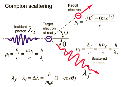
Compton Scattering
Key characteristics of Compton Scattering
Compton scattering dominates at intermediate energies.
It is the scattering of photons by atomic electrons
Photons undergo a wavelength shift called the Compton shift.
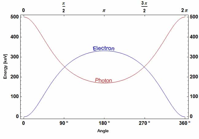
The energy transferred to the recoil electron can vary from zero to a large fraction of the incident gamma ray energy
Definition of Compton Scattering
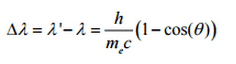
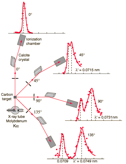
Compton scattering is the inelastic or nonclassical scattering of a photon (which may be an X-ray or gamma ray photon) by a charged particle, usually an electron. In Compton scattering, the incident gamma ray photon is deflected through an angle Θ with respect to its original direction. This deflection results in a decrease in energy (decrease in photon’s frequency) of the photon and is called the Compton effect. The photon transfers a portion of its energy to the recoil electron. The energy transferred to the recoil electron can vary from zero to a large fraction of the incident gamma ray energy, because all angles of scattering are possible. The Compton scattering was observed by A. H.Compton in 1923 at Washington University in St. Louis. Compton earned the Nobel Prize in Physics in 1927 for this new understanding about the particle-nature of photons.
Compton Scattering Formula
The Compton formula was published in 1923 in the Physical Review. Compton explained that the X-ray shift is caused by particle-like momentum of photons. Compton scattering formula is the mathematical relationship between the shift in wavelength and the scattering angle of the X-rays. In the case of Compton scattering the photon of frequency f collides with an electron at rest. Upon collision, the photon bounces off electron, giving up some of its initial energy (given by Planck’s formula E=hf), While the electron gains momentum (mass x velocity), the photon cannot lower its velocity. As a result of momentum conservetion law, the photon must lower its momentum given by:
So the decrease in photon’s momentum must be translated into decrease in frequency (increase in wavelength Δλ = λ’ – λ). The shift of the wavelength increased with scattering angle according to the Compton formula:
In Compton scattering, the incident gamma-ray photon is deflected through an angle Θ with respect to its original direction. This deflection results in a decrease in energy (decrease in photon’s frequency) of the photon and is called the Compton effect. Source: hyperphysics.phy-astr.gsu.edu
where
λ is the initial wavelength of photon
λ’ is the wavelength after scattering,
h is the Planck constant = 6.626 x 10⁻³⁴ J.s
me is the electron rest mass (0.511 MeV)
c is the speed of light
Θ is the scattering angle.
The minimum change in wavelength (λ′ − λ) for the photon occurs when Θ = 0° (cos(Θ)=1) and is at least zero. The maximum change in wavelength (λ′ − λ) for the photon occurs when Θ = 180° (cos(Θ)=-1). In this case the photon transfers to the electron as much momentum as possible.The maximum change in wavelength can be derived from Compton formula:
The quantity h/mec is known as the Compton wavelength of the electron and is equal to 2.43×10⁻¹² m.
Compton Scattering – Cross-Sections
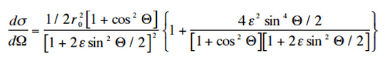
The probability of Compton scattering per one interaction with an atom increases linearly with atomic number Z, because it depends on the number of electrons, which are available for scattering in the target atom. The angular distribution of photons scattered from a single free electron is described by the Klein-Nishina formula:
where ε = E0/mec2 and r0 is the “classical radius of the electron” equal to about 2.8 x 10⁻¹³ cm. The formula gives the probability of scattering a photon into the solid angle element dΩ = 2π sin Θ dΘ when the incident energy is E0.
The wavelength change in such scattering depends only upon the angle of scattering for a given target particle. Source: hyperphysics.phy-astr.gsu.edu/
Cross section of compton scattering of photons by atomic electrons.Energies of a photon at 500 keV and an electron after Compton scattering. Source: wikipedia.org
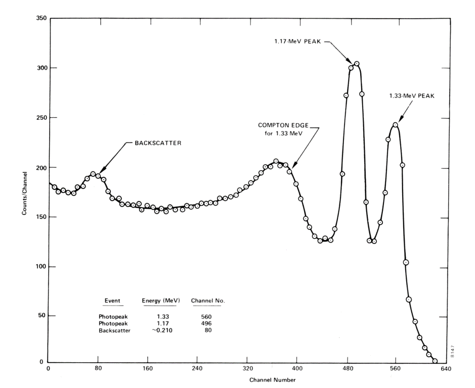
Compton Edge
In spectrophotometry, the Compton edge is a feature of the spectrograph that results from the Compton scattering in the scintillator or detector. This feature is due to photons that undergo Compton scattering with a scattering angle of 180° and then escape the detector. When a gamma ray scatters off the detector and escapes, only a fraction of its initial energy can be deposited in the sensitive layer of the detector. It depends on the scattering angle of the photon, how much energy will be deposited in the detector. This leads to a spectrum of energies. The Compton edge energy corresponds to full backscattered photon.
Inverse Compton Scattering
Inverse Compton scattering is the scattering of low energy photons to high energies by relativistic electrons. Relativistic electrons can boost energy of low energy photons by a potentially enormous amount (even gamma rays can be produced). This phenomenon is very important in astrophysics.
Compton edge of 60Co on gamma spectrometer Na(Tl).source: venables.asu.edu
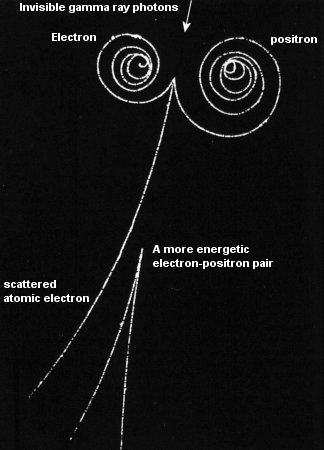
Positron-Electron Pair Production
In general, pair production is a phenomenon of nature where energy is direct converted to matter. The phenomenon of pair production can be view two different ways. One way is as a particle and antiparticle and the other is as a particle and a hole. The first way can be represented by formation of electron and positron, from a packet of electromagnetic energy (high energy photon – gamma ray) traveling through matter. It is one of the possible ways in which gamma rays interact with matter. At high energies this interaction dominates.
In order for electron-positron pair production to occur, the electromagnetic energy of the photon must be above a threshold energy, which is equivalent to the rest mass of two electrons. The threshold energy (the total rest mass of produced particles) for electron-positron pair production is equal to 1.02MeV (2 x 0.511MeV) because the rest mass of a single electron is equivalent to 0.511MeV of energy.
If the original photon’s energy is greater than 1.02MeV, any energy above 1.02MeV is according to the conservation law split between the kinetic energy of motion of the two particles.
The presence of an electric field of a heavy atom such as lead or uranium is essential in order to satisfy conservation of momentum and energy. In order to satisfy both conservation of momentum and energy, the atomic nucleus must receive some momentum. Therefore a photon pair production in free space cannot occur.
Moreover, the positron is the anti-particle of the electron, so when a positron comes to rest, it interacts with another electron, resulting in the annihilation of the both particles and the complete conversion of their rest mass back to pure energy (according to the E=mc2 formula) in the form of two oppositely directed 0.511 MeV gamma rays (photons). The pair production phenomenon is therefore connected with creation and destruction of matter in one reaction.
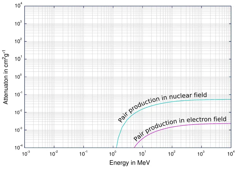
Positron-Electron Pair Production – Cross-Section
The probability of pair production, characterized by cross section, is a very complicated function based on quantum mechanics. In general the cross section increases approximately with the square of atomic number (σp ~ Z2) and increases with photon energy, but this dependence is much more complex.
Cross section of pair production in nuclear field and electron field.
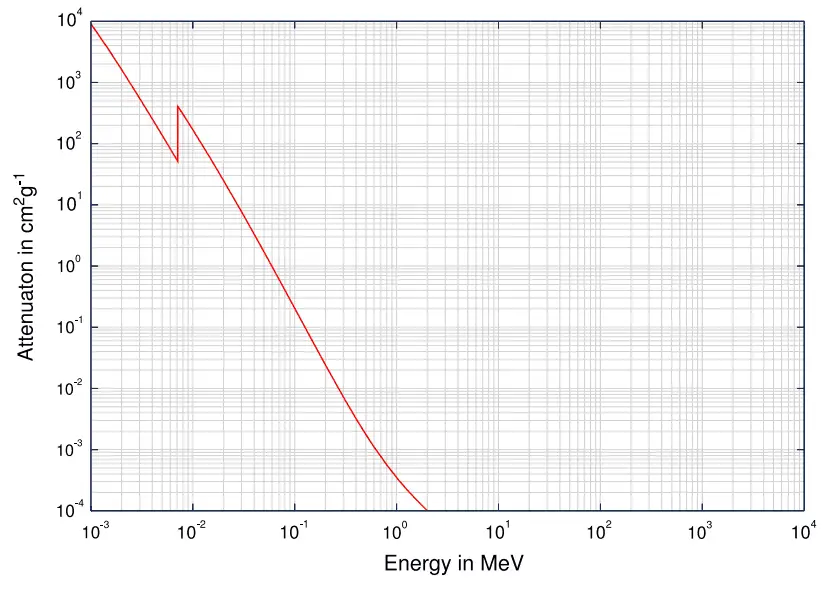
Gamma Rays Attenuation
The total cross-section of interaction of a gamma rays with an atom is equal to the sum of all three mentioned partial cross-sections:
σ = σf + σC + σp
σf – Photoelectric effect
σC – Compton scattering
σp – Pair production
Depending on the gamma ray energy and the absorber material, one of the three partial cross-sections may become much larger than the other two. At small values of gamma ray energy the photoelectric effect dominates. Compton scattering dominates at intermediate energies. The compton scattering also increases with decreasing atomic number of matter, therefore the interval of domination is wider for light nuclei. Finally, electron-positron pair production dominates at high energies.
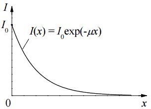
Based on the definition of interaction cross-section, the dependence of gamma rays intensity on thickness of absorber material can be derive. If monoenergetic gamma rays are collimated into a narrow beam and if the detector behind the material only detects the gamma rays that passed through that material without any kind of interaction with this material, then the dependence should be simple exponential attenuation of gamma rays. Each of these interactions removes the photon from the beam either by absorbtion or by scattering away from the detector direction. Therefore the interactions can be characterized by a fixed probability of occurance per unit path length in the absorber. The sum of these probabilities is called the linear attenuation coefficient:
μ = τ(photoelectric) + σ(Compton) + κ(pair)
The relative importance of various processes of gamma radiation interaction with matter.
Linear Attenuation Coefficient
The attenuation of gamma radiation can be then described by the following equation.
I=I0.e-μx
, where I is intensity after attenuation, Io is incident intensity, μ is the linear attenuation coefficient (cm-1), and physical thickness of absorber (cm).
Dependence of gamma radiation intensity on absorber thickness
The materials listed in the table beside are air, water and a different elements from carbon (Z=6) through to lead (Z=82) and their linear attenuation coefficients are given for three gamma ray energies. There are two main features of the linear attenuation coefficient:
The linear attenuation coefficient increases as the atomic number of the absorber increases.
The linear attenuation coefficient for all materials decreases with the energy of the gamma rays.
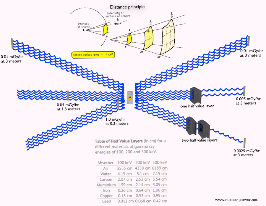
Half Value Layer
The half value layer expresses the thickness of absorbing material needed for reduction of the incident radiation intensity by a factor of two. There are two main features of the half value layer:
The half value layer decreases as the atomic number of the absorber increases. For example 35 m of air is needed to reduce the intensity of a 100 keV gamma ray beam by a factor of two whereas just 0.12 mm of lead can do the same thing.
The half value layer for all materials increases with the energy of the gamma rays. For example from 0.26 cm for iron at 100 keV to about 1.06 cm at 500 keV.
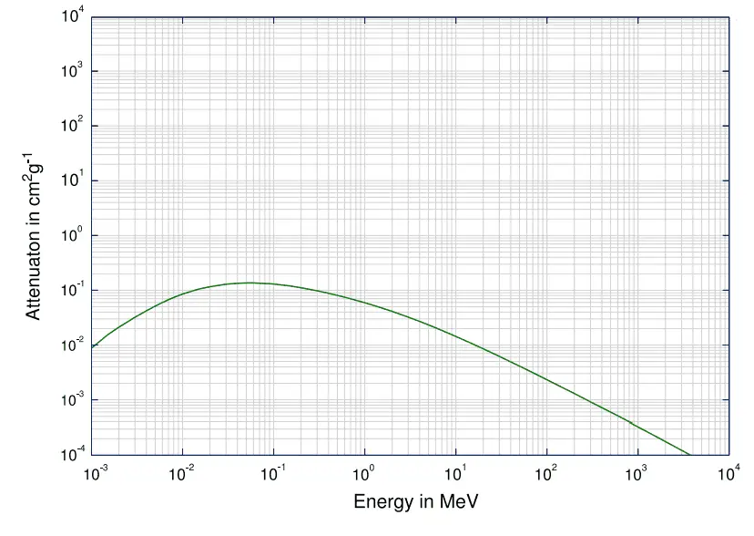
Mass Attenuation Coefficient
When characterizing an absorbing material, we can use sometimes the mass attenuation coefficient. The mass attenuation coefficient is defined as the ratio of the linear attenuation coefficient and absorber density (μ/ρ). The attenuation of gamma radiation can be then described by the following equation:
I=I0.e-(μ/ρ).ρl
, where ρ is the material density, (μ/ρ) is the mass attenuation coefficient and ρ.l is the mass thickness. The measurement unit used for the mass attenuation coefficient cm2g-1.
For intermediate energies the Compton scattering dominates and different absorbers have approximately equal mass attenuation coefficients. This is due to the fact that cross section of Compton scattering is proportional to the Z (atomic number) and therefore the coefficient is proportional to the material density ρ. At small values of gamma ray energy or at high values of gamma ray energy, where the coefficient is proportional to higher powers of the atomic number Z (for photoelectric effect σf ~ Z5; for pair production σp ~ Z2), the attenuation coefficient μ is not a constant.
Example:
How much water schielding do you require, if you want to reduce the intensity of a 500 keV monoenergetic gamma ray beam (narrow beam) to 1% of its incident intensity? The half value layer for 500 keV gamma rays in water is 7.15 cm and the linear attenuation coefficient for 500 keV gamma rays in water is 0.097 cm-1.
The question is quite simple and can be described by following equation:
If the half value layer for water is 7.15 cm, the linear attenuation coefficient is:
Now we can use the exponential attenuation equation:
therefore
So the required thickness of water is about 47.5 cm. This is relatively large thickness and it is caused by small atomic numbers of hydrogen and oxygen. If we calculate the same problem for lead (Pb), we obtain the thickness x=2.8cm.
Linear Attenuation Coefficients
Table of Linear Attenuation Coefficients (in cm-1) for a different materials at gamma ray energies of 100, 200 and 500 keV.
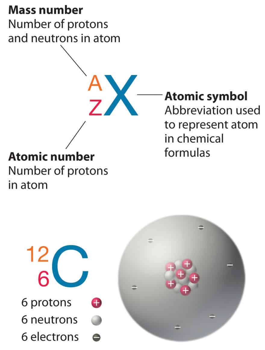
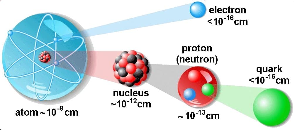
Atomic and Nuclear Structure. The atom consist of a small but massive nucleus surrounded by a cloud of rapidly moving electrons. The nucleus is composed of protons and neutrons. Periodic Table
Notation of nuclei Source: chemwiki.ucdavis.edu
The atom consist of a small but massive nucleus surrounded by a cloud of rapidly moving electrons. The nucleus is composed of protons and neutrons. Total number of protons in the nucleus is called the atomic number of the atom and is given the symbol Z. The total electrical charge of the nucleus is therefore +Ze, where e (elementary charge) equals to 1,602 x 10-19 coulombs. In a neutral atom there are as many electrons as protons moving about nucleus. It is the electrons that are responsible for the chemical bavavior of atoms, and which identify the various chemical elements.
Hydrogen (H), for example , consist of one electron and one proton. The number of neutrons in a nucleus is known as the neutron number and is given the symbol N. The total number of nucleons, that is, protons and neutrons in a nucleus, is equal to Z + N = A, where A is called the atomic mass number. The various species of atoms whose nuclei contain particular numbers of protons and neutrons are called nuclides. Each nuclide is denoted by chemical symbol of the element (this specifies Z) with tha atomic mass number as supescript.
Thus the symbol 1H refers to the nuclide of hydrogen with a single proton as nucleus. 2H is the hydrogen nuclide with a neutron as well as a proton in the nucleus (2H is also called deuterium or heavy hydrogen). Atoms such as 1H, 2H whose nuclei contain the same number of protons but different number of neutrons (different A) are known as isotopes. Uranium, for instance, has three isotopes occuring in nature – 238U, 235U and 234U. The stable isotopes (plus a few of the unstable isotopes) are the atoms that are found in the naturally occuring elements in nature. However, they are not found in equal amounts. Some isotopes of a given element are more abundant than others. For example 99,27% of naturally occuring uranium atoms are the isotope 238U, 0,72% are the isotope 235U and 0,0055% are the isotope 234U. Exact structure of atoms is described by Atomic Theory and Theory of Nuclear Structure.
Volume of an Atom and Nucleus
Structure of Matter.
The atom consist of a small but massive nucleus surrounded by a cloud of rapidly moving electrons. The nucleus is composed of protons and neutrons. Typical nuclear radii are of the order 10−14 m. Assuming spherical shape, nuclear radii can be calculated according to following formula:
r = r0 . A1/3
where r0 = 1.2 x 10⁻¹⁵ m = 1.2 fm
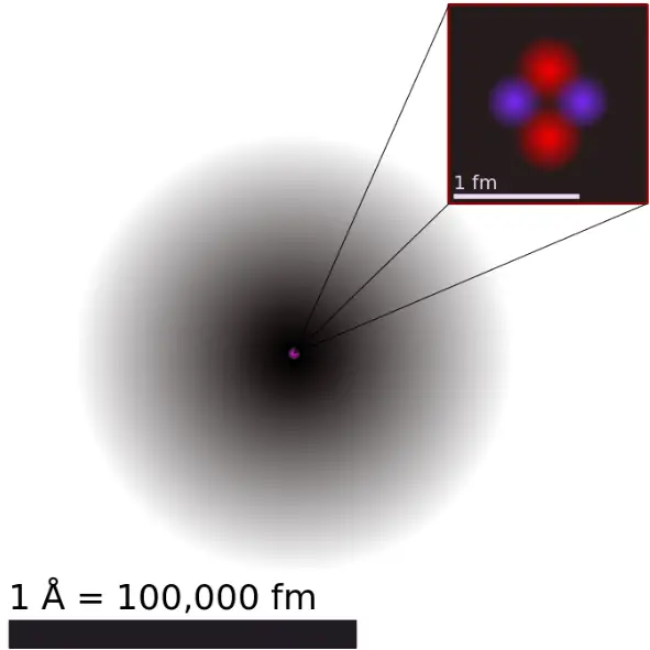
If we use this approximation, we therefore expect the volume of the nucleus to be of the order of 4/3πr3 or 7,23 ×10−45 m3 for hydrogen nuclei or 1721×10−45 m3 for 238U nuclei. These are volumes of nuclei and atomic nuclei (protons and neutrons) contains of about 99.95% of mass of atom.
Is an atom an empty space?
A figurative depiction of the helium-4 atom with the electron cloud in shades of gray. Protons and neutrons are most likely found in exactly the same space, at the central point. Source wikipedia.org License CC BY-SA 3.0
The volume of an atom is about 15 orders of magnitudelarger than the volume of a nucleus. For uranium atom, the Van der Waals radius is about 186 pm = 1.86 ×10⁻¹⁰ m. The Van der Waals radius, rw, of an atom is the radius of an imaginary hard sphere representing the distance of closest approach for another atom. Assuming spherical shape, the uranium atom have volume of about 26.9 ×10⁻³⁰ m3. But this “huge” space is occupied primarily by electrons, because the nucleus occupies only about 1721×10−45 m3 of space. These electrons together weigh only a fraction (let say 0.05%) of entire atom.
It may seem, that the space and in fact the matter is empty, but it is not. Due to the quantum nature of electrons, the electrons are not point particles, they are smeared out over the whole atom. The classical description cannot be used to describe things on the atomic scale. On the atomic scale, physicists have found that quantum mechanics describes things very well on that scale. Particle locations in quantum mechanics are not at an exact position, they are described by a probability density function. Therefore the space in an atom (between electrons and an atomic nucleus) is not empty, but it is filled by a probability density function of electrons (usually known as “electron cloud“).
Atomic Theory. Atomic theory is a scientific theory of the nature of matter, which states that matter is composed of discrete units called atoms. The word atom comes from the Ancient Greek adjective atomos, meaning “uncuttable”. Today it is known that also atoms are divisible. Atomic Theory consist of many models and discoveries, which gradually formed this theory.
Theory of Nuclear Structure. Understanding the structure of the atomic nucleus is one of the central challenges in modern nuclear physics.
Structure of Matter.
See also:
Fundamental Particles
See also:
Atomic and Nuclear Physics
See also:
Mass and Energy
We hope, this article, Atomic and Nuclear Structure, helps you. If so, give us a like in the sidebar. Main purpose of this website is to help the public to learn some interesting and important information about radiation and dosimeters.
What is radiation? How is radiation defined? Radiation is energy that comes from a source and travels through some material or through space. Light, heat and sound are types of radiation. Periodic Table
What is Radiation
Most general definition is that radiation is energy that comes from a source and travels through some material or through space. Light, heat and sound are types of radiation. This is very general definition, the kind of radiation discussed in this article is called ionizing radiation. Most people connect the term radiation only with ionizing radiation, but it is not correct. Radiation is all around us. In, around, and above the world we live in. It is a natural energy force that surrounds us. It is a part of our natural world that has been here since the birth of our planet. We should distinguish between:
Non-ionizing radiation. The kinetic energy of particles (photons, electrons, etc.) of non-ionizing radiation is too small to produce charged ions when passing through matter. The particles (photons) have only sufficient energy to change the rotational, vibrational or electronic valence configurations of target molecules and atoms. Sunlight, radio waves, and cell phone signals are examples of non-ionizing (photon) radiation. However, it can still cause harm, like when you get a sunburn.
Ionizing radiation. The kinetic energy of particles (photons, electrons, etc.) of ionizing radiation is sufficient and the particle can ionize (to form ion by losing electrons) target atoms to form ions. Simply ionizing radiation can knock electrons from an atom.
The boundary is not sharply defined, since different molecules and atoms ionize at different energies. This is typical for electromagnetic waves. Among electromagnetic waves belong, in order of increasing frequency (energy) and decreasing wavelength: radio waves, microwaves, infrared radiation, visible light, ultraviolet radiation, X-rays and gamma rays. Gamma rays, X-rays, and the higher ultraviolet part of the spectrum are ionizing, whereas the lower ultraviolet, visible light (including laser light), infrared, microwaves, and radio waves are considered non-ionizing radiation.
Forms of ionizing radiation
Interaction of Radiation with Matter
Ionizing radiation is categorized by the nature of the particles or electromagnetic waves that create the ionizing effect. These particles/waves have different ionization mechanisms, and may be grouped as:
Directly ionizing. Charged particles (atomic nuclei, electrons, positrons, protons, muons, etc.) can ionize atoms directly by fundamental interaction through the Coulomb force if it carries sufficient kinetic energy. These particles must be moving at relativistic speeds to reach the required kinetic energy. Even photons (gamma rays and X-rays) can ionize atoms directly (despite they are electrically neutral) through the Photoelectric effect and the Compton effect, but secondary (indirect) ionization is much more significant.
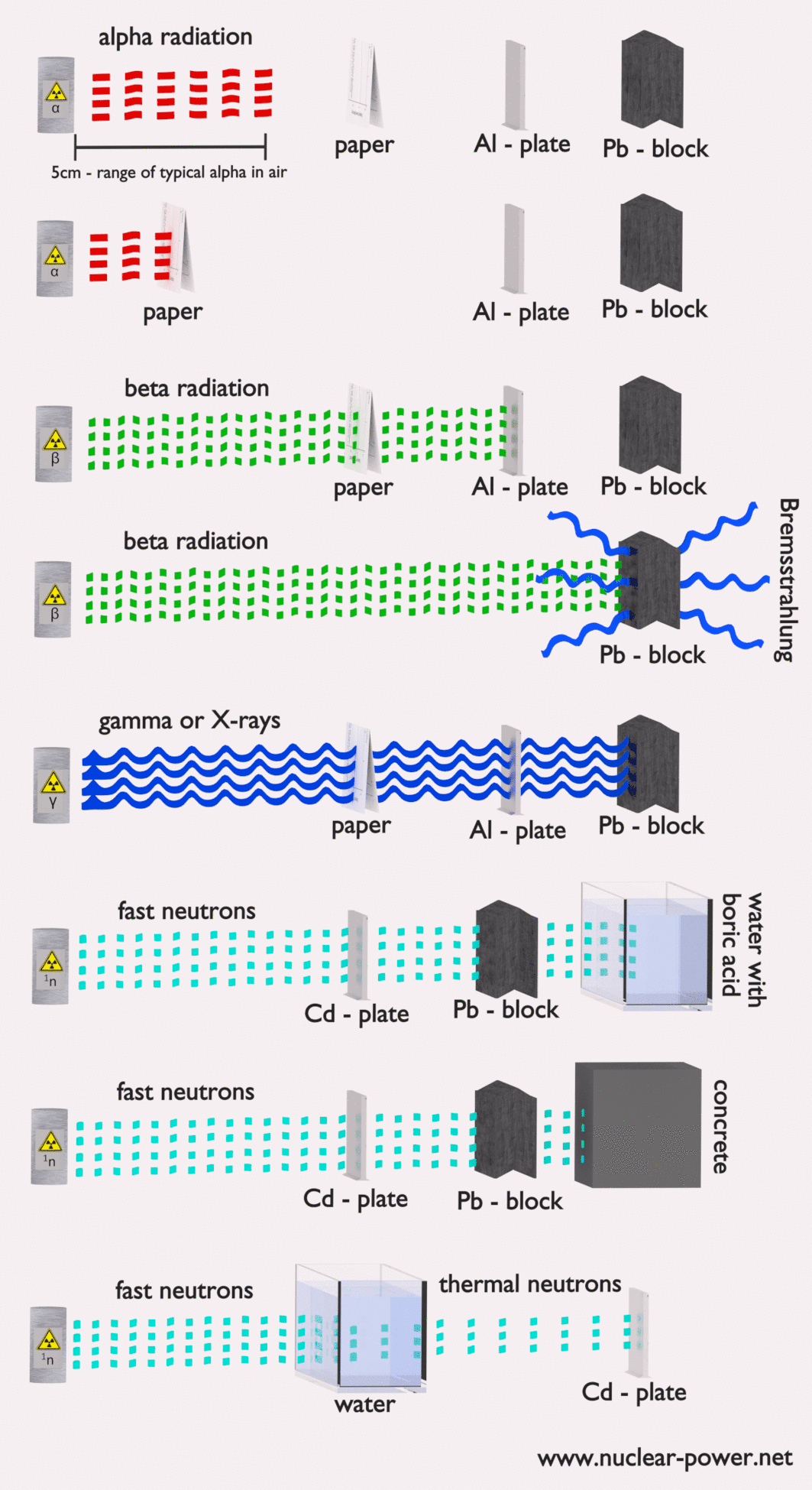
Alpha radiation. Alpha radiation consist of alpha particles at high energy/speed. The production of alpha particles is termed alpha decay. Alpha particles consist of two protons and two neutrons bound together into a particle identical to a helium nucleus. Alpha particles are relatively large and carry a double positive charge. They are not very penetrating and a piece of paper can stop them. They travel only a few centimeters but deposit all their energies along their short paths.
Beta radiation. Beta radiation consist of free electrons or positrons at relativistic speeds. Beta particles (electrons) are much smaller than alpha particles. They carry a single negative charge. They are more penetrating than alpha particles, but thin aluminum metal can stop them. They can travel several meters but deposit less energy at any one point along their paths than alpha particles.
Indirectly ionizing. Indirect ionizing radiation is electrically neutral particles and therefore does not interact strongly with matter. The bulk of the ionization effects are due to secondary ionizations.
Photon radiation (Gamma rays or X-rays). Photon radiation consist of high energy photons. These photons are particles/waves (Wave-Particle Duality) without rest mass or electrical charge. They can travel 10 meters or more in air. This is a long distance compared to alpha or beta particles. However, gamma rays deposit less energy along their paths. Lead, water, and concrete stop gamma radiation. Photons (gamma rays and X-rays) can ionize atoms directly through the Photoelectric effect and the Compton effect, where the relatively energetic electron is produced. The secondary electron will go on to produce multiple ionization events, therefore the secondary (indirect) ionization is much more significant.
Neutron radiation. Neutron radiation consist of free neutrons at any energies/speeds. Neutrons can be emitted by nuclear fission or by the decay of some radioactive atoms. Neutrons have zero electrical charge and cannot directly cause ionization. Neutrons ionize matter only indirectly. For example, when neutrons strike the hydrogen nuclei, proton radiation (fast protons) results. Neutrons can range from high speed, high energy particles to low speed, low energy particles (called thermal neutrons). Neutrons can travel hundreds of feet in air without any interaction.
Shielding of Ionizing Radiation
Radiation shielding simply means having some material between the source of radiation and you (or some device) that will absorb the radiation. The amount of shielding required, the type or material of shielding strongly depends on several factors. We are not talking about any optimisation.
In fact in some cases an inappropriate shielding may even worsen the radiation situation instead of protecting people from the ionizing radiation. Basic factors, which have to be considered during proposal of radiation shielding, are:
Type of the ionizing radiation to be shielded
Energy spectrum of the ionizing radiation
Length of exposure
Distance from the source of the ionizing radiation
Requirements on the attenuation of the ionizing radiation – ALARA or ALARP principles
Design degree of freedom
Other physical requirements (e.g. transparence in case of leaded glass screens)
Generally in nuclear industry the radiation shielding has many purposes. In nuclear power plants the main purpose is to reduce the radiation exposure to persons and staff in the vicinity of radiation sources. In NPPs the main source of radiation is conclusively the nuclear reactor and its reactor core. Nuclear reactors are in generall powerful sources of entire spectrum of types of ionizing radiation. Shielding used for this purpose is called biological shielding.
But this is not the only purpose of radiation shielding. Shields are also used in some reactors to reduce the intensity of gamma rays or neutrons incident on the reactor vessel. This radiation shielding protects the reactor vessel and its internals (e.g. the core support barrel) from the excessive heating due to gamma ray absorption fast neutron moderation. Such shields are usually referred to as thermal shields.
A little strange radiation shielding is usually used to protect material of reactor pressure vessel (especially in PWR power plants). Structural materials of pressure vessel and reactor internals are damaged especially by fast neutrons. Fast neutrons create structural defects, which in result lead to embrittlement of material of pressure vessel. In order to minimize the neutron flux at the vessel wall, also core loading strategy can be modified. In “out-in” fuel loading strategy fresh fuel assemblies are placed at the periphery of the core. This configuration causes high neutron fluence at the vessel wall. Therefore the “in-out” fuel loading strategy (with low leakage loading patterns – L3P) has been adopted at many nuclear power plants. In contrast to “out-in” strategy, low leakage cores have fresh fuel assemblies in the second row, not at the periphery of the core. The periphery contains fuel with higher fuel burnup and lower relative power and serves as the very sophisticated radiation shield.
In nuclear power plants the central problem is to shield against gamma rays and neutrons, because the ranges of charged particles (such as beta particles and alpha particles) in matter are very short. On the other hand we must deal with shielding of all types of radiation, because each nuclear reactor is a significant source of all types of ionizing radiation.
See also:
Mass and Energy
See also:
Atomic and Nuclear Physics
See also:
Nuclear Stability
We hope, this article, Radiation, helps you. If so, give us a like in the sidebar. Main purpose of this website is to help the public to learn some interesting and important information about radiation and dosimeters.
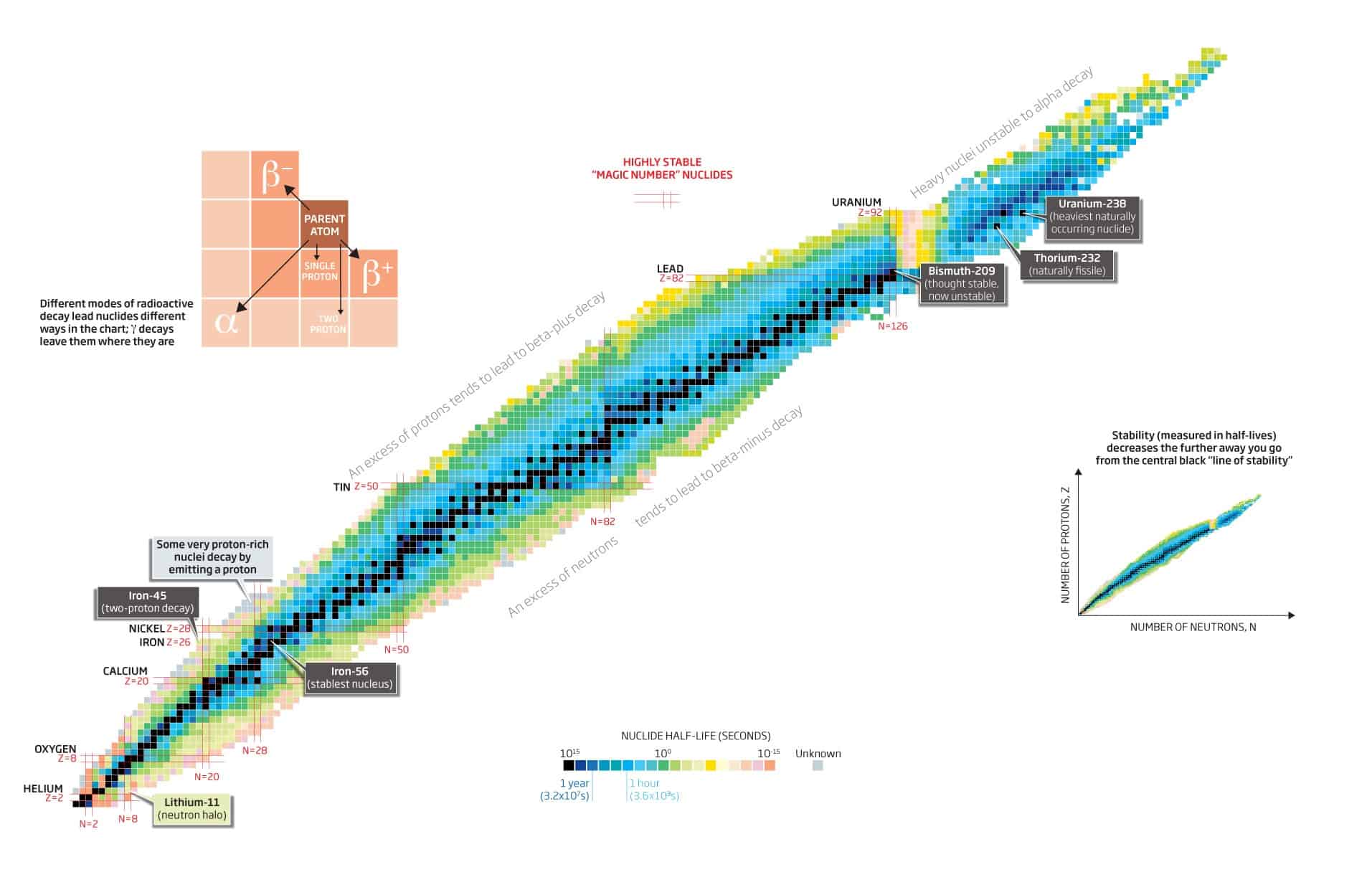
Nuclear Stability is a concept that helps to identify the stability of an isotope. To identify the stability of an isotope it is needed to find the ratio of neutrons to protons. Periodic Table
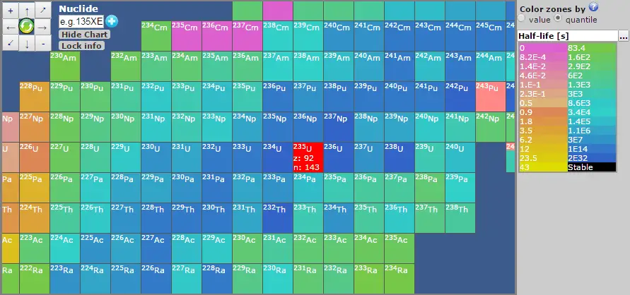
Nuclear Stability is a concept that helps to identify the stability of an isotope. To identify the stability of an isotope it is needed to find the ratio of neutrons to protons. To determine the stability of an isotope you can use the ratio neutron/proton (N/Z). Also to help understand this concept there is a chart of the nuclides, known as a Segre chart. This chart shows a plot of the known nuclides as a function of their atomic and neutron numbers. It can be observed from the chart that there are more neutrons than protons in nuclides with Z greater than about 20 (Calcium). These extra neutrons are necessary for stability of the heavier nuclei. The excess neutrons act somewhat like nuclear glue.
Detail of Nuclide Chart. Source: Livechart – IAEA.org
Atomic nuclei consist of protons and neutrons, which attract each other through the nuclear force, while protons repel each other via the electric force due to their positive charge. These two forces compete, leading to various stability of nuclei. There are only certain combinations of neutrons and protons, which forms stable nuclei.
Neutrons stabilize the nucleus, because they attract each other and protons , which helps offset the electrical repulsion between protons. As a result, as the number of protons increases, an increasing ratio of neutrons to protons is needed to form a stable nucleus. If there are too many or too few neutrons for a given number of protons, the resulting nucleus is not stable and it undergoes radioactive decay. Unstable isotopes decay through various radioactive decay pathways, most commonly alpha decay, beta decay, or electron capture. Many other rare types of decay, such as spontaneous fission or neutron emission are known. It should be noted that all of these decay pathways may be accompanied by the subsequent emission of gamma radiation. Pure alpha or beta decays are very rare.
Examples:
Positive beta decay
Nuclei, such as 15O, which are lacking in neutrons (consist of 8 protons and 7 neutrons) undergo positron decay (positive beta decay). In this process, one of the protons in the nucleus is transformed into a neutron, positron and neutrino.The positron and the neutrino are emitted. The number of protons is thus reduced from 8 to 7 (number of neutrons is increased from 7 to 8), so that the resulting nucleus is an isotope of nitrogen, 15N, which is stable.
Negative beta decay
On the other hand nuclei, such as 19O, which have excess of neutrons, decay by negative beta decay, emitting a negative electron and an antineutrino. In this process, one of the neutrons in the nucleus is transformed into a proton. The number of protons is thus increased from 8 to 9 (number of neutrons is reduced from 11 to 10), so that the resulting nucleus is an isotope of fluor, 19F, which is stable. It should be noted that in both positive or negative beta decays the atomic mass number remains the same.
Nuclear Stability – Periodic Table
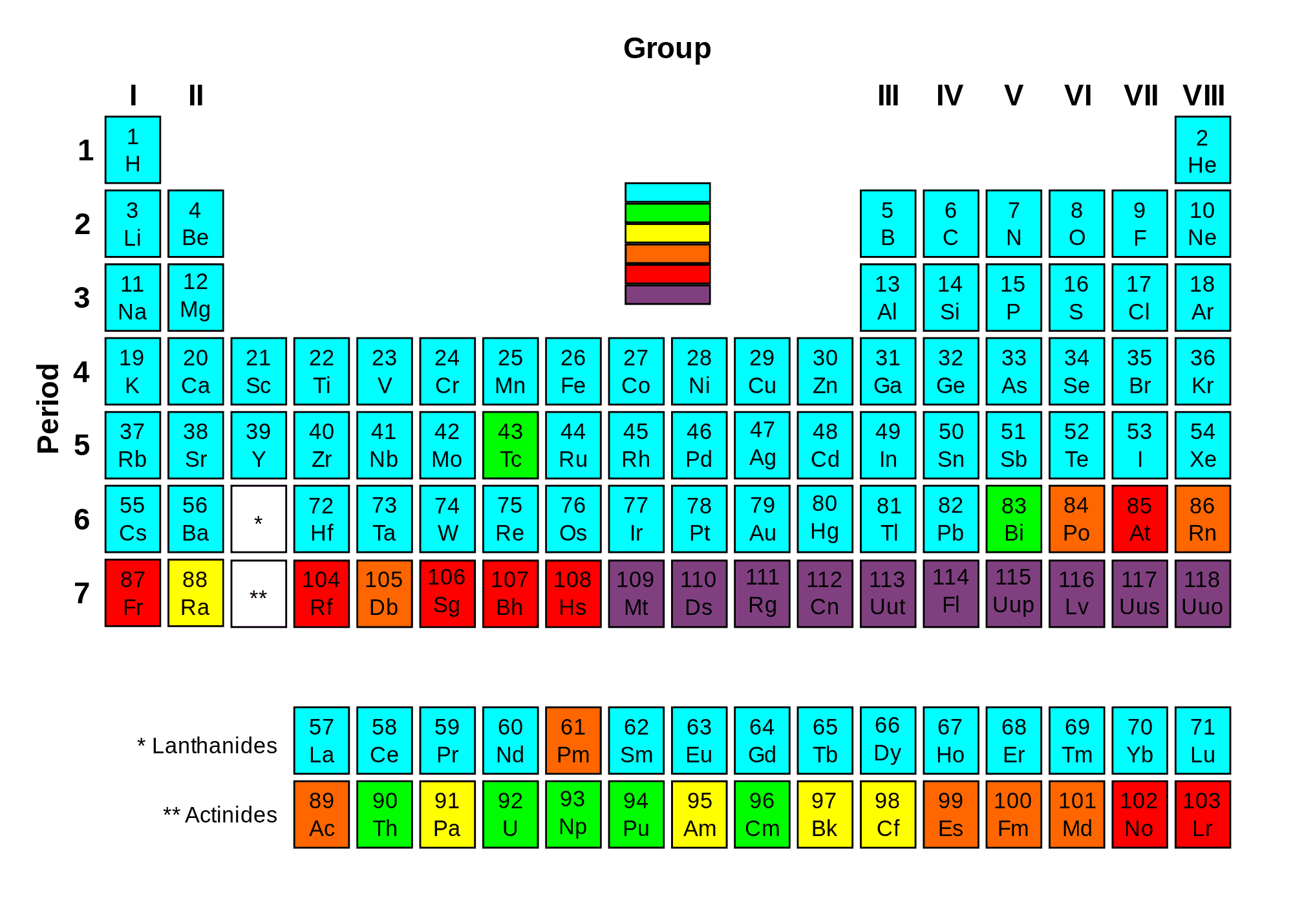
Periodic table with elements colored according to the half-life of their most stable isotope.
Of the first 82 elements in the periodic table, 80 have isotopes considered to be stable. Technetium, promethium and all the elements with an atomic number over 82 are unstable and decompose through radioactive decay. No undiscovered heavy elements (with atomic number over 110) are expected to be stable, therefore lead is considered the heaviest stable element. For each of the 80 stable elements, the number of the stable isotopes is given. For example, tin has 10 such stable isotopes.
There are 80 elements with at least one stable isotope, but 114 to 118 chemical elements are known. All elements to element 98 are found in nature, and the remainder of the discovered elements are artificially produced, with isotopes all known to be highly radioactive with relatively short half-lives.
Bismuth, thorium, uranium and plutonium are primordial nuclides because they have half-lives long enough to still be found on the Earth, while all the others are produced either by radioactive decay or are synthesized in laboratories and nuclear reactors. Primordial nuclides are nuclides found on the Earth that have existed in their current form since before Earth was formed. Primordial nuclides are residues from the Big Bang, from cosmogenic sources, and from ancient supernova explosions which occurred before the formation of the solar system. Only 288 such nuclides are known.
Connection between Nuclear Stability and Radioactive Decay
The nuclei of radioisotopes are unstable. In an attempt to reach a more stable arrangement of its neutrons and protons, the unstable nucleus will spontaneously decay to form a different nucleus. If the number of neutrons changes in the process (number of protons remains), a different isotopes is formed and an element remains (e.g. neutron emission). If the number of protons changes (different atomic number) in the process, then an atom of a different element is formed. This decomposition of the nucleus is referred to as radioactive decay. During radioactive decay an unstable nucleus spontaneosly and randomly decomposes to form a different nucleus (or a different energy state – gamma decay), giving off radiation in the form of atomic partices or high energy rays. This decay occurs at a constant, predictable rate that is referred to as half-life. A stable nucleus will not undergo this kind of decay and is thus non-radioactive.
See also:
Radiation
See also:
Atomic and Nuclear Physics
See also:
Radioactive Decay
We hope, this article, Nuclear Stability, helps you. If so, give us a like in the sidebar. Main purpose of this website is to help the public to learn some interesting and important information about radiation and dosimeters.
The neutron was discovered in 1932 by the English physicist James Chadwick. The story of the discovery of the neutron is central to the extraordinary developments in atomic physics. Periodic Table
The story of the discovery of the neutron and its properties is central to the extraordinary developments in atomic physics that occurred in the first half of the 20th century. The neutron was discovered in 1932 by the English physicist James Chadwick, but since the time of Ernest Rutherford it had been known that the atomic mass number A of nuclei is a bit more than twice the atomic number Z for most atoms and that essentially all the mass of the atom is concentrated in the relatively tiny nucleus. The Rutherford’s model for the atom in 1911 claims that atoms have their mass and positive charge concentrated in a very small nucleus.
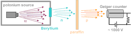
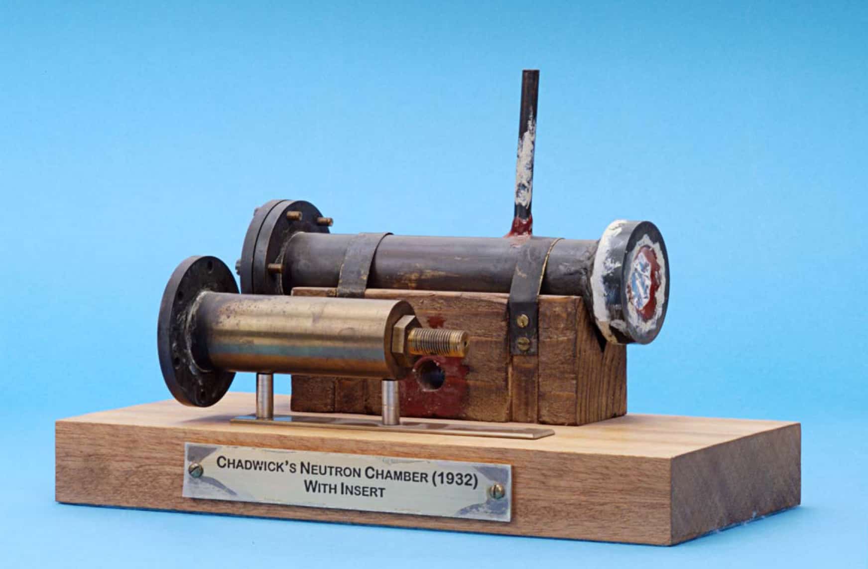
The alpha particles emitted from polonium fell on certain light elements, specifically beryllium, an unusually penetrating radiation is produced. Source: dev.physicslab.orgChadwick’s neutron chamber containing parallel disks of radioactive polonium and beryllium. Radiation is emitted from an aluminium window at the chamber’s end. Source: imgkid.com
An experimental breakthrough came in 1930 with the observation by Bothe and Becker. They found that if the very energetic alpha particles emitted from polonium fell on certain light elements, specifically beryllium, boron, or lithium, an unusually penetrating radiation was produced. Since this radiation was not influenced by an electric field (neutrons have no charge), they presumed it was gamma rays (but much more penetrating). It was shown (Curie and Joliot) that when a paraffin target with this radiation is bombarded, it ejected protons with energy about 5.3 MeV. Paraffin is high in hydrogen content, hence offers a target dense with protons (since neutrons and protons have almost equal mass, protons scatter energetically from neutrons).These experimental results were difficult to interpret. James Chadwick was able to prove that the neutral particle could not be a photon by bombarding targets other than hydrogen, including nitrogen, oxygen, helium and argon. Not only were these inconsistent with photon emission on energy grounds, the cross-section for the interactions was orders of magnitude greater than that for Compton scattering by photons. In Rome, the young physicist Ettore Majorana suggested that the manner in which the new radiation interacted with protons required a new neutral particle.
The task was that of determining the mass of this neutral particle. James Chadwick chose to bombard boron with alpha particles and analyze the interaction of the neutral particles with nitrogen. These particlular targets were chosen partly because the masses of boron and nitrogen were well known. Using kinematics, Chadwick was able to determine the velocity of the protons. Then through conservation of momentum techniques, he was able to determine that the mass of the neutral radiation was almost exactly the same as that of a proton. In 1932, Chadwick proposed that the neutral particle was Rutherford’s neutron. In 1935, he was awarded the Nobel Prize for his discovery.
See also:
Neutron
See also:
Structure of the Neutron
We hope, this article, Discovery of the Neutron, helps you. If so, give us a like in the sidebar. Main purpose of this website is to help the public to learn some interesting and important information about radiation and dosimeters.
The quark structure of the neutron. Forces between quarks in neutrons are mediated by gluons. Neutrons and protons are classified as hadrons and baryons. Periodic Table
The quark structure of the neutron. The color assignment of individual quarks is arbitrary, but all three colors must be present. Forces between quarks are mediated by gluons.
Neutrons and protons are classified as hadrons, subatomic particles that are subject to the strong force and as baryons since they are composed of three quarks. The neutron is a composite particle made of two down quarks with charge −⅓ e and one up quark with charge +⅔ e. Since the neutron has no net electric charge, it is not affected by eletric forces, but the neutron does have a slight distribution of electric charge within it. This results in non-zero magnetic moment (dipole moment) of the neutron. Therefore the neutron interacts also via electromagnetic interaction, but much weaker than the proton.
The mass of the neutron is 939.565 MeV/c2, whereas the mass of the three quarks is only about 12 MeV/c2 (only about 1% of the mass-energy of the neutron). Like the proton, most of mass (energy) of the neutron is in the form of the strong nuclear force energy (gluons). The quarks of the neutron are held together by gluons, the exchange particles for the strong nuclear force. Gluons carry the color charge of the strong nuclear force.
See also:
Discovery of the Neutron
See also:
Neutron
See also:
Properties of the Neutron
We hope, this article, Structure of the Neutron, helps you. If so, give us a like in the sidebar. Main purpose of this website is to help the public to learn some interesting and important information about radiation and dosimeters.
Neutrons may interact with matter in many ways. Neutrons are neutral particles,therefore they collide with nuclei, not with atoms. Interactions of Neutrons with Matter. Periodic Table
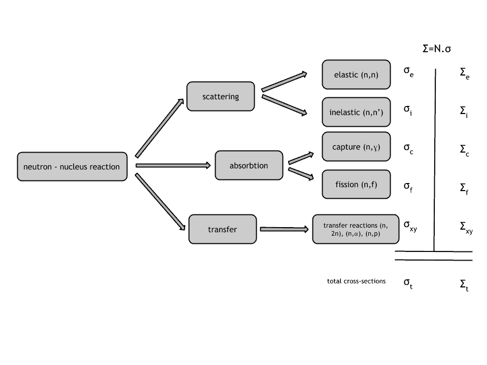
Interactions of Neutrons with Matter
Neutrons are neutral particles, therefore they travel in straight lines, deviating from their path only when they actually collide with a nucleus to be scattered into a new direction or absorbed. Neither the electrons surrounding (atomic electron cloud) a nucleus nor the electric field caused by a positively charged nucleus affect a neutron’s flight. In short, neutrons collide with nuclei, not with atoms. A very descriptive feature of the transmission of neutrons through bulk matter is the mean free path length (λ – lambda), which is the mean distance a neutron travels between interactions. It can be calculated from following equation:
λ=1/Σ
Neutrons may interact with nuclei in one of following ways:
Types of neutron-nuclear reactions
Elastic Scattering Reaction
Generally, a neutron scattering reaction occurs when a target nucleus emits a single neutron after a neutron-nucleus interaction. In an elastic scattering reaction between a neutron and a target nucleus, there is no energy transferred into nuclear excitation.
Inelastic Scattering Reaction
In an inelastic scattering reaction between a neutron and a target nucleus some energy of the incident neutron is absorbed to the recoiling nucleus and the nucleus remains in the excited state. Thus while momentum is conserved in an inelastic collision, kinetic energy of the “system” is not conserved.
Neutron Absorption
The neutron absorption reaction is the most important type of reactions that take place in a nuclear reactor. The absorption reactions are reactions, where the neutron is completely absorbed and compound nucleus is formed. This is the very important feature, because the mode of decay of such compound nucleus does not depend on the way the compound nucleus was formed. Therefore a variety of emissions or decays may follow. The most important absorption reactions are divided by the exit channel into two following reactions:
Radiative Capture. Most absorption reactions result in the loss of a neutron coupled with the production of one or more gamma rays. This is referred to as a capture reaction, and it is denoted by σγ.
Neutron-induced Fission Reaction. Some nuclei (fissionable nuclei) may undergo a fission event, leading to two or more fission fragments (nuclei of intermediate atomic weight) and a few neutrons. In a fissionable material, the neutron may simply be captured, or it may cause nuclear fission. For fissionable materials we thus divide the absorption cross section as σa = σγ + σf.
Radiative Capture
The neutron capture is one of the possible absorption reactions that may occur. In fact, for non-fissionable nuclei it is the only possible absorption reaction. Capture reactions result in the loss of a neutron coupled with the production of one or more gamma rays. This capture reaction is also referred to as a radiative capture or (n, γ) reaction, and its cross-section is denoted by σγ.
The radiative capture is a reaction, in which the incident neutron is completely absorbed and compound nucleus is formed. The compound nucleus then decays to its ground state by gamma emission. This process can occur at all incident neutron energies, but the probability of the interaction strongly depends on the incident neutron energy and also on the target energy (temperature). In fact the energy in the center-of-mass system determines this probability.
Nuclear Fission
Nuclear fission is a nuclear reaction in which the nucleus of an atom splits into smaller parts (lighter nuclei). The fission process often produces free neutrons and photons (in the form of gamma rays), and releases a large amount of energy. In nuclear physics, nuclear fission is either a nuclear reactionor a radioactive decay process. The case of decay process is called spontaneous fission and it is very rare process.
Neutron Emission
Although the neutron emission is usually associated with nuclear decay, it must be also mentioned in connection with neutron nuclear reactions. Some neutrons interacts with a target nucleus via a compound nucleus. Among these compound nucleus reactions are also reactions, in which a neutron is ejected from nucleus and they may be referred to as neutron emission reactions. The point is that compound nuclei lose its excitation energy in a way, which is identical to the radioactive decay. Very important feature is the fact the mode of decay of compound nucleus does not depend on the way the compound nucleus was formed.
Charged Particle Ejection
Charged particle reactionsare usually associated with formation of a compound nucleus, which is excited to a high energy level, that such compound nucleus can eject a new charged particle while the incident neutron remains in the nucleus. After the new particle is ejected, the remaining nucleus is completely changed, but may or may not exist in an excited state depending upon the mass-energy balance of the reaction. This type of reaction is more common for charged particles as incident particles (such as alpha particles, protons, and so on).
The case of neutron-induced charged particle reactions is not so common, but there are some neutron-induced charged particle reactions, that are of importance in the reactivity control and also in the detection of neutrons.
Neutron cross-section
Typical cross-sections of fission material. Slowing down neutrons results in increase of probability of interaction (e.g. fission reaction).
The extent to which neutrons interact with nuclei is described in terms of quantities known as cross-sections. Cross-sections are used to express the likelihood of particular interaction between an incident neutron and a target nucleus. It must be noted this likelihood do not depend on real target dimensions. In conjunction with the neutron flux, it enables the calculation of the reaction rate, for example to derive the thermal power of a nuclear power plant. The standard unit for measuring the microscopic cross-section (σ-sigma) is the barn, which is equal to 10-28 m2. This unit is very small, therefore barns (abbreviated as “b”) are commonly used. The microscopic cross-section can be interpreted as the effective ‘target area’ that a nucleus interacts with an incident neutron.
A macroscopic cross-section is derived from microscopic and the material density:
Σ=σ.N
Here σ, which has units of m2, is referred to as the microscopic cross-section. Since the units of N (nuclei density) are nuclei/m3, the macroscopic cross-sectionΣ have units of m-1, thus in fact is an incorrect name, because it is not a correct unit of cross-sections.
Neutron cross-sections constitute a key parameters of nuclear fuel. Neutron cross-sections must be calculated for fresh fuel assemblies usually in two-Dimensional models of the fuel lattice.
The neutron cross-section is variable and depends on:
Target nucleus (hydrogen, boron, uranium, etc.) Each isotop has its own set of cross-sections.
Type of the reaction (capture, fission, etc.). Cross-sections are different for each nuclear reaction.
Neutron energy (thermal neutron, resonance neutron, fast neutron). For a given target and reaction type, the cross-section is strongly dependent on the neutron energy. In the common case, the cross section is usually much larger at low energies than at high energies. This is why most nuclear reactors use a neutron moderator to reduce the energy of the neutron and thus increase the probability of fission, essential to produce energy and sustain the chain reaction.
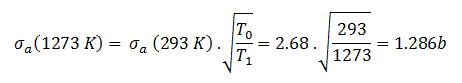
Target energy (temperature of target material – Doppler broadening) This dependency is not so significant, but the target energy strongly influences inherent safety of nuclear reactors due to a Doppler broadening of resonances.
For thermal neutrons (in 1/v region), absorption cross sections increases as the velocity (kinetic energy) of the neutron decreases. Source: JANIS 4.0
For thermal neutrons (in 1/v region), absorption cross-sections increases as the velocity (kinetic energy) of the neutron decreases. Therefore the 1/v Law can be used to determine shift in absorbtion cross-section, if the neutron is in equilibrium with a surrounding medium. This phenomenon is due to the fact the nuclear force between the target nucleus and the neutron has a longer time to interact.
This law is aplicable only for absorbtion cross-section and only in the 1/v region.
Example of cross- sections in 1/v region:
The absorbtion cross-section for 238U at 20°C = 293K (~0.0253 eV) is:
.
The absorbtion cross-section for 238U at 1000°C = 1273K is equal to:
This cross-section reduction is caused only due to the shift of temperature of surrounding medium.
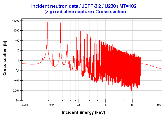
Resonance neutron capture
Resonance peaks for radiative capture of U238. At resonance energies the probability of capture can be more than 100x higher as the base value. Source: JANIS program
Absorption cross section is often highly dependent on neutron energy. Note that the nuclear fission produces neutrons with a mean energy of 2 MeV (200 TJ/kg, i.e. 20,000 km/s). The neutron can be roughly divided into three energy ranges:
Fast neutron. (10MeV – 1keV)
Resonance neutron (1keV – 1eV)
Thermal neutron. (1eV – 0.025eV)
The resonance neutrons are called resonance for their special bahavior. At resonance energies the cross-section can reach peaks more than 100x higher as the base value of cross-section. At this energies the neutron capture significantly exceeds a probability of fission. Therefore it is very important (for thermal reactors) to quicklyovercome this range of energy and operate the reactor with thermal neutrons resulting in increase of probability of fission.
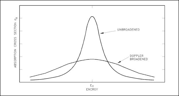
Doppler broadening
Doppler effect improves reactor stability. Broadened resonance (heating of a fuel) results in a higher probability of absorbtion, thus causes negative reactivity insertion (reduction of reactor power).
A Doppler broadening of resonances is very important phanomenon, which improves reactor stability. The prompt temperature coefficient of most thermal reactors is negative, owing to an nuclear Doppler effect. Although the absorbtion cross-section depends significantly on incident neutron energy, the shape of the cross-section curve depends also on target temperature.
Nuclei are located in atoms which are themselves in continual motion owing to their thermal energy. As a result of these thermal motions neutrons impinging on a target appears to the nuclei in the target to have a continuous spread in energy. This, in turn, has an effect on the observed shape of resonance. The resonance becomes shorter and wider than when the nuclei are at rest.
Although the shape of a resonance changes with temperature, the total area under the resonance remains essentially constant. But this does not implyconstant neutron absorbtion. Despite the constant area under resonance, a resonance integral, which determines the absorbtion, increases with increasing target temperature. This, of course, decreases coefficient k (negative reactivity is inserted).
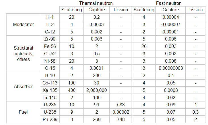
Typical cross-sections of materials in the reactor
Following table shows neutron cross-sections of the most common isotopes of reactor core.
Table of cross-sections
See also:
Neutron Energy
See also:
Neutron
See also:
Free Neutron
We hope, this article, Interaction of Neutrons with Matter, helps you. If so, give us a like in the sidebar. Main purpose of this website is to help the public to learn some interesting and important information about radiation and dosimeters.
We use cookies to ensure that we give you the best experience on our website. If you continue to use this site we will assume that you are happy with it.Ok